August 2026 ECHO Question of the Month Authors:
Amber McCranie, MS4
UT Southwestern Medical Center
Ingrid Moreno-Duarte, MD
Assistant Professor of Anesthesiology
Program Director for the Pediatric Cardiac Anesthesiology Fellowship
Department of Anesthesiology and Pain Management
Divisions of Adult and Pediatric Cardiothoracic Anesthesiology and Adult Critical Care Medicine
University of Texas Southwestern / Children’s Medical Center Dallas
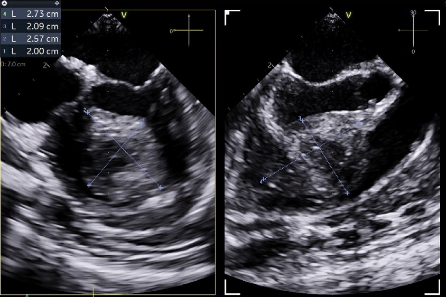
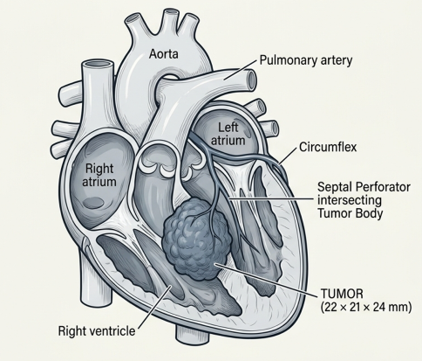
A 14-month-old male, born at 29 weeks’ gestation with a small muscular ventricular septal defect (VSD) was followed with serial transthoracic echocardiography (TTE) for VSD surveillance. At 6 months of age, surveillance TTE incidentally identified a small, approximately 1 mm mass within the interventricular septum. The previous muscular VSD had spontaneously closed. Repeat echocardiography 6 months later demonstrated substantial interval growth to 15×17 mm. Cardiac MRI at an outside hospital demonstrated a 17×18×17 mm mid-interventricular septal mass with imaging characteristics consistent with a cardiac fibroma. Subsequent cardiac CT (Image 1) for further anatomic characterization and surgical planning demonstrated a 22×21×24 mm mass centered within the interventricular septum, with predominant extension toward the right ventricle (RV) and right ventricular outflow tract (RVOT). Biventricular systolic function was preserved, coronary artery anatomy was normal, and a septal perforator arising from the left anterior descending coronary artery was seen coursing toward the mass. The patient remained asymptomatic, without arrhythmias, syncope, cyanosis, or signs of heart failure. Given the rapid interval growth of the mass, surgical resection was planned.
Image 1
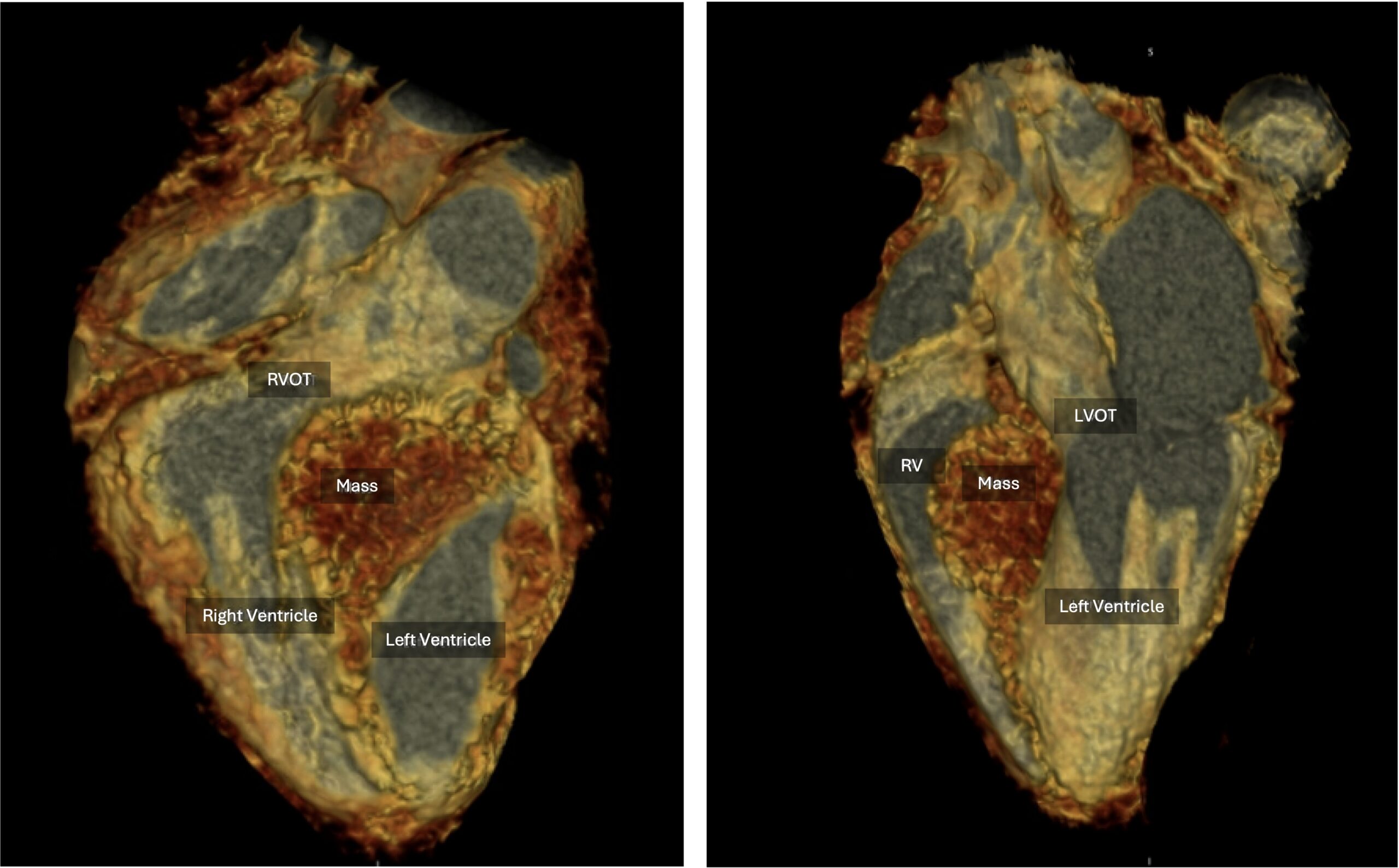
Image 2
Discussion:
Cardiac fibromas are the second most common benign pediatric cardiac tumor after rhabdomyomas and, unlike rhabdomyomas, do not typically regress spontaneously.[4] They are composed predominantly of fibroblasts and collagen and generally present as solitary, well-circumscribed intramyocardial masses.[2,4] Cardiac fibromas most commonly arise within the ventricular myocardium, particularly the left ventricular free wall or interventricular septum, and less frequently involve the right ventricle.[1,4]
Depending on their size and location, cardiac fibromas may result in ventricular outflow tract obstruction, arrhythmias, conduction abnormalities, or compression of adjacent coronary arteries.[2–4] Clinical presentation is variable, ranging from an incidental finding in an asymptomatic patient to murmur, syncope, heart failure, ventricular arrhythmias, or sudden cardiac death.[2–4] Importantly, cardiac fibroma and larger tumor dimensions have been associated with an increased risk of malignant ventricular arrhythmias and cardiovascular death in children.[3]
Surgical management may involve complete excision or subtotal resection when complete removal would compromise adjacent myocardium or other critical cardiac structures. [1,2] Resection of large or deeply intramyocardial tumors may require ventricular or septal reconstruction, including patch closure of a resulting ventricular septal defect. Particular concerns with resection of interventricular septal fibromas include injury to the conduction system and compromise of adjacent coronary arterial branches. In addition, surgical resection may result in regional wall-motion abnormalities and reduced ventricular systolic function; larger tumor volume has been associated with greater postoperative regional wall-motion abnormalities, and postoperative left ventricular dysfunction has been described following resection of left ventricular fibromas.[1]
This patient remained asymptomatic despite substantial interval growth of the mass and progressive protrusion into the right ventricle and right ventricular outflow tract, prompting surgical resection. Multimodality imaging with transthoracic echocardiography, cardiac MRI, cardiac CT, and perioperative transesophageal echocardiography allowed serial assessment of tumor growth, characterization of its location and tissue features, delineation of its relationship to the coronary circulation and surrounding cardiac structures, and assessment of its hemodynamic impact and ventricular function.[2,4]
For a more in-depth discussion on the echo exam for cardiac fibroma, please view the echo tutorial here.
The pre-operative transesophageal echocardiographic assessment should include the following:
- Biventricular cavity size and function
- Mass size and location
- Mass extension and impact on nearby structures
- Presence and degree of RVOT and LVOT obstruction
- Evaluation of AV valve function
- Coronary artery anatomy
- Additional VSDs, if present
- Pericardial effusion
Post-operative echocardiographic assessment should focus on the following
- Biventricular cavity size and systolic function
- Residual VSD or free wall defects
- Aneurysms at or near repair sites
References:
- Beroukhim RS, Geva T, Del Nido P, et al. Risk Factors for Left Ventricular Dysfunction Following Surgical Management of Cardiac Fibroma. Circ Cardiovasc Imaging. 2021 Feb;14(2):e011748. doi: 10.1161/CIRCIMAGING.120.011748. Epub 2021 Feb 1. PMID: 33517672.
- Covington MK, Young PM, Bois MC, et al. Clinical Impact of Cardiac Fibromas. Am J Cardiol. 2022 Nov 1;182:95-103. doi: 10.1016/j.amjcard.2022.06.062. Epub 2022 Aug 30. PMID: 36055811.
- Placidi S, Calcagni G, Lioncino M, et al. Type and dimensions can predict ventricular arrhythmias and cardiac death in primary benign cardiac tumors in children. Int J Cardiol. 2025 Jan 1;418:132599. doi: 10.1016/j.ijcard.2024.132599. Epub 2024 Sep 24. PMID: 39326704.
- Tao TY, Yahyavi-Firouz-Abadi N, Singh GK, Bhalla S. Pediatric cardiac tumors: clinical and imaging features. 2014 Jul-Aug;34(4):1031-46. doi: 10.1148/rg.344135163. PMID: 25019440.