September 2026 ECHO Question of the Month Authors:
Emma Xi Li, MD, FASE, FACC
Assistant Professor
Department of Anesthesiology and Perioperative Medicine
Mayo Clinic Rochester
Vy An Tran, MD, MSc
Assistant Professor
Pediatric Cardiac Anesthesiology
Department of Anesthesiology, Critical Care and Pain Medicine
The University of Texas Health Science Center at Houston
Question 1
What type of ASD is shown in Video 1 above? Which type of ASD is amenable to transcatheter closure, and which rims are assessed?
Answer
Video 1 is a superior septal view with color flow doppler. It shows the RA, LA and atrial septum with a large secundum ASD. Typically, only the ostium secundum ASD is amenable to percutaneous closure, while sinus venosus, primum, and coronary sinus defects require surgical closure.1 In selected cases, superior sinus venosus ASDs can be closed with covered stents. 2 However, this is a newer approach and is not the standard of practice at most institutions.

Image 1: Secundum ASD rims and surrounding structures. Image generated by OpenAI, ChatGPT-5.6Sol, 2026
For secundum ASDs, by convention there are six rims: aortic (superior/anterior, also called the retro-aortic rim), atrioventricular (inferior/anterior), superior vena cava (SVC), inferior vena cava (IVC), posterior, and right upper pulmonary vein rims.2 A rim length of ≥5 mm is generally considered favorable; a rim <5 mm is termed "deficient." Adequate posteroinferior rims—particularly the IVC rim—are most critical for a stable device landing zone, and absence of the IVC rim is considered a contraindication to device closure.1 A deficient aortic rim is not necessarily an absolute contraindication but shifts device selection toward a softer occluder and heightens post-deployment vigilance.
Question 2
Using intracardiac echocardiography (ICE) with the catheter in the right atrium, which imaging view best profiles the superior vena cava (SVC) and inferior vena cava (IVC) rims?
Answer
With the phased-array ICE catheter positioned in the right atrium, the septum is interrogated systematically through a set of standard views that mirror the corresponding TEE planes:
- Home view (tricuspid/RV) at 12 o’clock: initial orientation with the tricuspid valve and right ventricle.
- Septal (long-axis of the interatrial septum) view: profiles the length of the septum and the atrioventricular and posterosuperior margins. Tilt the catheter posteriorly to sweep across the interatrial septum.
- Long-axis (caval) view: flex the anterior-posterior knob cephalad or caudal to bring in the SVC or IVC, profiling the SVC and IVC rims. Use slight clockwise or counter-clockwise motion to further fine-tune these rims
- Short-axis (aortic) view at 4:30-5:00 o’clock: further clockwise rotation to profile the aortic (retro-aortic) and posterior rims.1,3,4
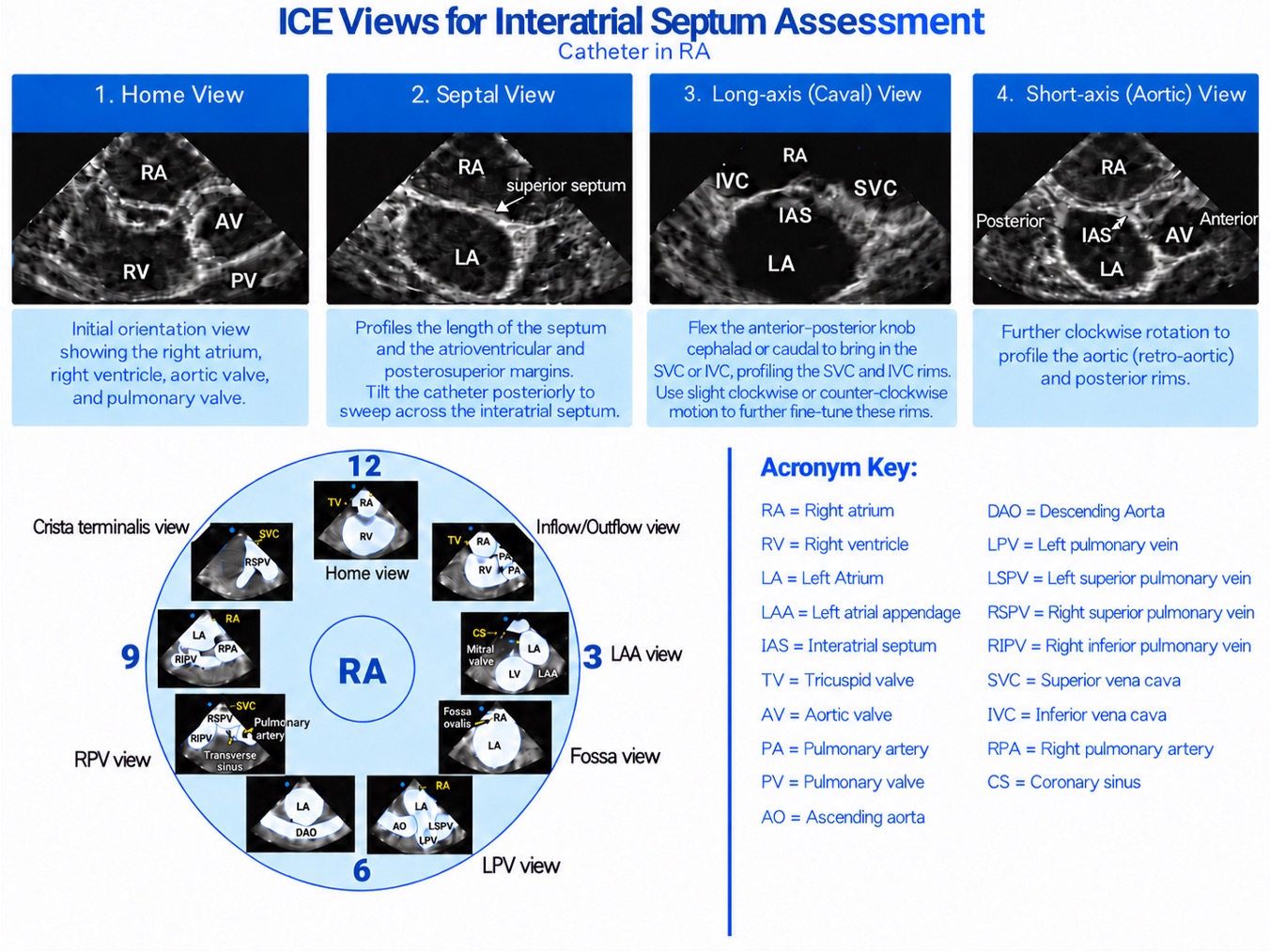
Image 2: Relevant interatrial septal views and standard clock view for ICE. Image generated by OpenAI, ChatGPT-5.6Sol, 2026.
A particular strength of ICE is superior visualization of the inferior/posterior rims that are frequently foreshortened on TEE, and 3D/multiplane (xPlane). ICE increasingly allows en-face and simultaneous biplane rim assessment analogous to 3D TEE.
Discussion:
Contemporary ASD Closure Devices
Transcatheter closure is becoming the standard of care for the majority of anatomically suitable secundum ASDs and is performed via femoral venous access under combined fluoroscopic and echocardiographic guidance. Most centers perform elective closure in children >15 kg and ≥3–5 years of age—our 15-year-old is well within this range. Three device families are currently FDA-approved:
- Amplatzer Septal Occluder (ASO; Abbott): approved in 2001, a self-centering, self-expanding double-disc device of braided nitinol filled with Dacron/polyester patches, with a central waist that sits within the defect. It closes defects up to ~38–40 mm and has the longest track record. Its main device-specific concern is erosion (0.1–0.3%), linked to oversizing and deficient retro-aortic rim; erosion rates have declined substantially since instructions-for-use updates addressing rim deficiency. 5,6
- Amplatzer Cribriform (Multi-Fenestrated) Occluder (Abbott): a non–self-centering device with a thin waist and equal discs, designed for multi-fenestrated septa/aneurysms.
- Gore Cardioform Septal Occluder (GCSO; W.L. Gore): a soft nitinol frame covered with expanded polytetrafluoroethylene (PTFE), non–self-centering, approved for secundum ASDs up to 17 mm (and for PFO).
- Gore Cardioform ASD Occluder (GCA; W.L. Gore): approved in 2019, a self-centering ePTFE/nitinol device with an adaptable central waist that conforms to the defect, closing defects ~8–35 mm. Its soft profile has been associated with essentially no reported erosions, though wire-frame fractures can occur.
- Occlutech ASD Occluder (B. Braun): approved in 2023, a self-expanding double disc system with various combinations of LA and RA disc sizes and waist dimensions
Device choice depends on defect size, morphology, rim adequacy, and operator preference. Newer bioresorbable occluders are under investigation in Europe but are not standard of care. Post-procedure, antiplatelet therapy (typically aspirin) for at least 6 months and endocarditis prophylaxis for 6 months are recommended.
This patient’s fluoroscopy images just before and after the final device release are shown below.
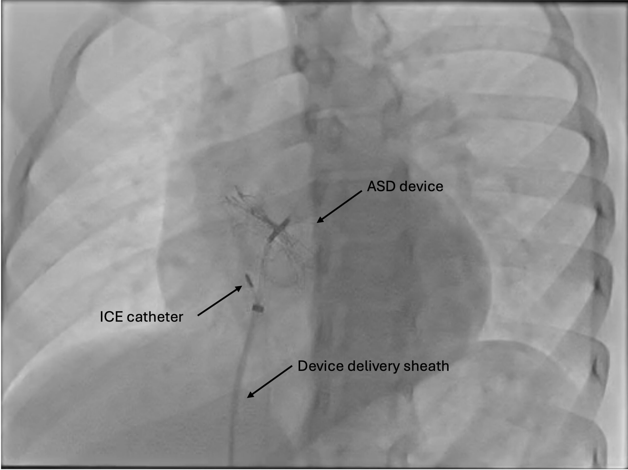
Image 3: Fluoroscopy image showing Gore Cardioform ASD occluder with delivery sheath and ICE catheter both inserted via the femoral vein sitting within the RA.
Video 4: Fluoroscopy showing deployed ASD device and contrast originating within SVC, distributing into the RA. Notably, there is no contrast seen passing into the LA, confirming a well seated ASD device.
Intracardiac Echocardiography (ICE)
ICE is an increasingly used alternative to TEE for guiding transcatheter ASD closure and carries specific implications for the anesthesia team. A phased-array catheter is advanced from a second femoral venous sheath into the right atrium, providing high-resolution imaging of the septum, rims, and device from within the heart. Standard ICE views—home (tricuspid/RV), septal (long-axis of the interatrial septum), long-axis (caval), and short-axis (aortic)—mirror the corresponding TEE planes and are combined with color Doppler and fluoroscopy.
The principal advantages are that ICE can be performed under conscious/moderate sedation rather than general anesthesia—avoiding an esophageal probe and airway instrumentation—and it often better visualizes the inferior/posterior rims that can be foreshortened on TEE, with reported reductions in fluoroscopy and procedure time. Trade-offs include the need for additional venous access, added cost of a single-use catheter, a largely single-plane right-atrial imaging perspective (though 3D/multiplane ICE is emerging), and the fact that the interventionalist typically drives the imaging.
In a 15-year-old, TEE under general anesthesia remains a common and excellent choice, but ICE (or a pediatric/micro-TEE probe) is a reasonable option, particularly to avoid deep sedation. From the anesthesiologist’s standpoint, ICE-based cases still require vigilance for vascular access complications, air embolism, and the rare acute complications (effusion, tamponade, embolization) that echocardiography is deployed to detect.
References:
- Faletra et al. “Imaging for Patient’s Selection and Guidance of LAA and ASD Percutaneous and Surgical Closure”. JACC Cardiovasc Imaging. 2021 Jan;14(1):3-21. doi: 10.1016/j.jcmg.2019.06.032.
- Baruteau AE, et al. “Transcatheter Closure of Superior Sinus Venosus Defects” JACC Cardiovasc Interv. 2023 Nov 13;16(21):2587-2599. doi: 10.1016/j.jcin.2023.07.024. Epub 2023 Oct 18. PMID: 37855807.
- Silvestry et al. “Guidelines for the Echocardiographic Assessment of Atrial Septal Defect and Patent Foramen Ovale: From the American Society of Echocardiography and Society for Cardiac Angiography and Interventions”. ASE Guidelines & Standards. Volume 28, Issue 8p910-958August 2015.
- Enriquez et al. “Use of Intracardiac Echocardiography in Interventional Cardiology: Working With the Anatomy Rather Than Fighting It”. Circulation. 2018 May 22;137(21):2278-2294. doi: 10.1161/CIRCULATIONAHA.117.031343.
- Turner et al. “Transcatheter Closure of Atrial and Ventricular Septal Defects: JACC Focus Seminar”. J Am Coll Cardiol. 2022 Jun 7;79(22):2247-2258. doi: 10.1016/j.jacc.2021.08.082.
- Levi DS, McElhinney DB. Long-Term Trends in Cardiac Erosion After Transcatheter Closure of Atrial Septal Defects With the Amplatzer Septal Occluder. Catheter Cardiovasc Interv. 2026 Sep;108(3):875-882. doi: 10.1002/ccd.70722. Epub 2026 Jul 6. PMID: 42403250; PMCID: PMC13532425.