Author: Anila B Elliott, MD - University of Michigan, C.S. Mott Children’s Hospital
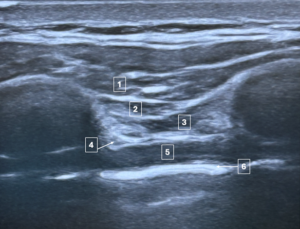
A 10-year-old with bicuspid aortic valve presents for a Ross procedure. They are consented for a deep parasternal intercostal plane (DPIP) block. What is the correct order of anatomical structures in the ultrasound image below, from superficial to deep (1 to 6)?
EXPLANATION
The deep parasternal intercostal plane (DPIP) block is a regional technique used to provide analgesia to the anterior chest wall. This block targets the anterior branches of the intercostal nerves (T2-T6) as they course anterior to the internal mammary vasculature. Local anesthetic is deposited into the fascial layer between the internal intercostal muscle and the transversus thoracic muscle1,2. As local anesthetic is injected, these layers are separated, as illustrated in Figure 1.
Figure 1: Image A: The needle is seen traversing the tissue layers prior to injection of local anesthetic. Note the location of the pleura (arrow). Image B: Following injection, the anesthetic spreads and separates the tissue layers, displacing the pleura inferiorly.

Utilization of the DPIP block has been shown to decrease post-operative pain scores and reduce opioid requirements following pediatric cardiac surgery3. Cadaveric studies have shown that the local anesthetic spread of the DPIP block covers a broader area than the superficial parasternal muscle plane (SPIP) block, offering enhanced pain control4. However, the DPIP is considered a more advanced block as it carries a potential risk of vascular injury, as the needle tip may come within millimeters of the internal mammary artery 4,5.
Other regional blocks of the anterior chest wall include the PECS I and PECS II blocks. The PECS I block traverses the pectoralis major and deposits local anesthetic between the pectoralis major and pectoralis minor, targeting the pectoral nerves. The PECS II block traverses the pectoralis major and pectoralis minor muscles to deposit local anesthetic between the pectoralis minor and the serratus anterior muscle, targeting the lateral cutaneous intercostal nerves (T2-T6)6.
The correct answer is A – the layers depicted in the picture from superficial to deep are the pectoralis major, external intercostal muscle, internal intercostal muscle, transversus thoracic muscle, internal mammary artery, and the pleura.
REFERENCES
1. Cakmak M, Isik O. Transversus Thoracic Muscle Plane Block for Analgesia After Pediatric Cardiac Surgery. J Cardiothorac Vasc Anesth. 2021;35(1):130-136. doi:10.1053/j.jvca.2020.07.053
2. Aydin ME, Ahiskalioglu A, Ates I, et al. Efficacy of Ultrasound-Guided Transversus Thoracic Muscle Plane Block on Postoperative Opioid Consumption After Cardiac Surgery: A Prospective, Randomized, Double-Blind Study. J Cardiothorac Vasc Anesth. 2020;34(11):2996-3003. doi:10.1053/j.jvca.2020.06.044
3. Cui YY, Xu ZQ, Hou HJ, Zhang J, Xue JJ. Transversus Thoracic Muscle Plane Block For Postoperative Pain in Pediatric Cardiac Surgery: A Systematic Review And Meta-Analysis of Randomized And Observational Studies. J Cardiothorac Vasc Anesth. 2024;38(5):1228-1238. doi:10.1053/j.jvca.2024.02.016
4. Douglas RN, Kattil P, Lachman N, et al. Superficial versus deep parasternal intercostal plane blocks: cadaveric evaluation of injectate spread. Br J Anaesth. 2024;132(5):1153-1159. doi:10.1016/j.bja.2023.08.014
5. Sepolvere G, Tognù A, Tedesco M, Coppolino F, Cristiano L. Avoiding the Internal Mammary Artery During Parasternal Blocks: Ultrasound Identification and Technique Considerations. J Cardiothorac Vasc Anesth. 2021;35(6):1594-1602. doi:10.1053/j.jvca.2020.11.007
6. Dost B, De Cassai A, Amaral S, et al. Regional anesthesia for pediatric cardiac surgery: a review. BMC Anesthesiol. 2025;25(1):77. Published 2025 Feb 15. doi:10.1186/s12871-025-02960-z