Authors: Kirsten Groody, MD AND Anila B. Elliott, MD - University of Michigan - C.S. Mott Children’s Hospital
A 6-month-old 5 kg male undergoes closure of a large, unrestrictive perimembranous ventricular septal defect (VSD). Post-repair transesophageal echo (TEE) shows a 4mm residual shunt. Assuming normal pulmonary vascular resistance (PVR) and no pulmonary venous desaturation, what values would you expect on a saturation run on an FiO2 of 21%?
Ventricular septal defects (VSD) are among the most common form of congenital heart disease. They are classified according to location in the ventricular septum, with perimembranous (also called conoventricular) defects being the most common subtype.
The pathophysiology of a VSD is characterized by a left-to-right shunt, resulting in a ratio of pulmonary blood flow to systemic blood flow (Qp:Qs) greater than one (Qp:Qs>1). The degree of shunting is dependent on the size of the VSD and the pulmonary vascular resistance (PVR): larger defects and lower PVR produce more left-to-right shunting and increase pulmonary blood flow (PBF). Persistent pulmonary over circulation increases volume and workload for the left ventricle, leading to LV dilation and congestive heart failure (CHF)1. These typically manifest as poor feeding, tachypnea, diaphoresis, and poor weight gain. Increased flow to the pulmonary vascular bed can also result in pulmonary vascular disease and pulmonary hypertension. If a VSD is left unrepaired, PVR may exceed systemic vascular resistance, eventually resulting in right-to-left shunting across the VSD and cyanosis (Eisenmenger syndrome)1.
Muscular defects are most likely to close spontaneously. In contrast, large perimembranous defects are less likely to close and often warrant surgical intervention. Timing for surgery is often around 6 months of age, though often guided by clinical signs of CHF and diagnostic findings on TTE. Cardiac catheterization is rarely indicated before surgery. If obtained, a Qp:Qs greater than 2:1 warrants intervention to prevent any progression of pulmonary vascular disease1.
Overall mortality from VSD repair is low (<1%), however residual VSDs are fairly common3. Potential risks associated with residual VSD include infection, continued shunt physiology, and valvular abnormalities. Intra-operative transesophageal echocardiography is useful to determine adequacy of repair. Defects less than 4mm are often physiologically insignificant, and defects less than 2mm often close spontaneously2. In cases where TEE results are equivocal or the residual jet is difficult to quantify, a “saturation run” can help determine whether the residual shunt is physiologically significant by estimating Qp:Qs. Qp:Qs is determined using the following equation, where SaO2 is systemic arterial oxygen saturation, SmvO2 is mixed venous oxygen saturation, SpvO2 is pulmonary venous oxygen saturation, and SpaO2 is pulmonary arterial oxygen saturation 3,4.
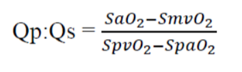
Superior vena cava (SVC) saturation best reflects mixed venous oxygen saturation in patients with open shunts and are best obtained on room air 4. Pulmonary venous saturation can be presumed to be roughly equal to aortic saturation, assuming healthy lungs. Typically, a Qp:Qs greater than 1.5-2:1 warrants consideration for return to cardiopulmonary bypass and would typically produce a pulmonary artery step-up above the mixed venous saturation. In our patient with a 4mm residual defect, using the above equation, answer A yields a Qp:Qs 1.8:1 with a step-up in PAO2, answer B a Qp:Qs 1:1, and answer C Qp:Qs 0.7:1. Therefore, the correct answer is A.
REFERENCES
1. Bradley, S. M. (2025). Ventricular Septal Defects (VSD). In Mokadam, N., Jacobs, J., & Meyerson, S. (Eds.), Adult and Pediatric Cardiac Surgery. STS Cardiothoracic Surgery E-Book. Chicago: Society of Thoracic Surgeons. ebook.sts.org
2. Bibevski S, Ruzmetov M, Mendoza L, et al. The Destiny of Postoperative Residual Ventricular Septal Defects After Surgical Repair in Infants and Children. World Journal for Pediatric and Congenital Heart Surgery 2020;11(4):438-443
3. Yang SG, Novello R, Nicolson S, Steven J, Gaynor JW, Spray TL, Rychik J. Evaluation of ventricular septal defect repair using intraoperative transesophageal echocardiography: frequency and significance of residual defects in infants and children. Echocardiography 2000;17(7):681-684
4. Stark, RJ; Shekerdemian, LS. Estimating intracardiac and extracardiac shunting in the setting of complex congenital heart disease. Annals of Pediatric Cardiology 2013;6(2):145-151