
Author: Manal Mirreh, MD - Children’s Hospital of Philadelphia
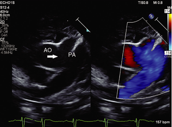
A 3-day-old neonate undergoes echocardiography for a murmur. Parasternal short-axis imaging (below) is obtained, with color Doppler demonstrating antegrade flow from the ascending aorta (AO) into the adjacent main pulmonary artery (PA), with both branch pulmonary arteries arising normally from the main PA. Which of the following lesions is shown in the echocardiogram?
EXPLANATION
The image shows a side-to-side communication between the ascending aorta and an otherwise normally-formed main pulmonary artery, with both branch pulmonary arteries arising appropriately from the main PA, which is the defining anatomy of an aortopulmonary window (APW) (Answer C).
Embryologically, an APW results from failure of the aortopulmonary septum to fuse with the neural crest-derived outflow cushions, leaving a persistent aortopulmonary foramen between two vessels that are otherwise fully and separately septated, each with its own semilunar valve.1
Aortopulmonary window is a rare anomaly comprising approximately 0.1-0.6% of all congenital heart defects. The basic anatomical defect in APW consists of communication between the ascending aorta and the pulmonary artery.
Mori and colleagues classified APW into three types (I–III); the STS classification later added a fourth, 'intermediate' type:
Type I: proximal defect located just above the sinus of Valsalva, a few millimeters above the semilunar valve
Type II: distal APW located in the uppermost portion of ascending aorta
Type III: total defect involving the majority of the ascending aorta
Type IV: an intermediate defect, classified as neither proximal nor distal
The pressure gradient between the aorta and pulmonary artery will produce significant left-to-right shunting depending on the size of the defect and the relative resistances of the pulmonary and systemic vascular beds.2 Surgical closure is therefore indicated in all patients with aortopulmonary window and it should be undertaken as early as possible after diagnosis because of the risk of pulmonary vascular disease.
Simple APW has been defined as without any significant associated anomalies, or anomalies requiring minor or simple repair (patent ductus arteriosus, atrial septal defect, patent foramen ovale). Complex APW is a defect occurring with more complex associated anomalies such as IAA, transposition, tetralogy of Fallot, or anomalous origin of the coronary arteries. The risk of morbidity and mortality in these patients is related to the associated cardiac defects, in particular the association of interrupted aortic arch.3 Uncorrected APW results in a reported 40% mortality in the first year of life with a substantial proportion of survivors succumbing to CHF later in childhood.2

From Backer CL, Mavroudis C. Surgical management of aortopulmonary window: a 40-year experience. Eur J Cardiothorac Surg. 2002;21:773–779.
Answer A, anomalous origin of the pulmonary artery from the aorta (AOPA), is incorrect. AOPA occurs due to failure of closure of the embryonic aortopulmonary foramen, resulting in a branch pulmonary artery arising directly from the ascending aorta rather than from the main pulmonary artery.
AOPA and APW do arise from the same underlying process, a deficiency in septation of the truncus arteriosus, and both have been described in terms of failure of closure of the embryonic aortopulmonary foramen. AOPA involves an entire branch pulmonary artery (usually the right) failing to align with its 6th arch-derived pulmonary channel, and instead arises directly from the ascending aorta. On echocardiogram, the main PA would connect to only one branch PA, with the other branch taking off from the aorta. In this image, both branch pulmonary arteries arise normally from the main PA, which rules out AOPA and points instead to a true AP window, a side-to-side communication between two vessels that are otherwise completely and separately formed.
Answer B, window ductus, is incorrect because it would be anatomically remote from what is shown on the displayed echocardiogram. A PDA is a 6th arch derivative connecting the main PA to the descending aorta, not the ascending aorta. Even a window-morphology (Krichenko type E) PDA remains, by definition, a duct-derived structure inserting on the descending aorta, so it would not produce the ascending aorta-to-PA color flow seen here.
REFERENCES
1. Mehta ID, Porayette P, Rivera RJ, Buddhavarapu A, Mehta C. Aortopulmonary Window; Hemitruncus. In: Critical Heart Disease in Infants and Children. 3rd ed. Elsevier; 2019:652-660.e1.
2. Andropoulos DB, Mossad EB, Gottlieb EA. Anesthesia for Left-to-Right Shunt Lesions. In: Anesthesia for Congenital Heart Disease 4th ed. Wiley; 2023:628-631.
3. C.L. Backer, C. Mavroudis, Surgical management of aortopulmonary window: a 40-year experience, European Journal of Cardio-Thoracic Surgery, Volume 21, Issue 5, May 2002, Pages 773–779, doi.org/10.1016/S1010-7940(02)00056-8