Congenital Cardiac Anesthesia Society
A Section of the Society for Pediatric Anesthesia.

ECHO Question of the Month – July 2026 – Keyword: Tetralogy of Fallot

July 2026 ECHO Question of the Month Author:
Vy An Tran, MD, MSc
Assistant Professor
Pediatric Cardiac Anesthesiology
Department of Anesthesiology, Critical Care and Pain Medicine
The University of Texas Health Science Center at Houston


An 11-month-old male, born at 36-weeks’ gestation with prenatal diagnosis of Tetralogy of Fallot (ToF) and multilevel pulmonary obstruction requiring PDA stenting at 2 weeks of age, presented for full repair. Pre-operative cardiac CT revealed an anomalous LAD arising from the RCA orifice, crossing the RV outflow tract at the level of the pulmonary valve annulus. The transesophageal echo (TEE) prior to cardiopulmonary bypass revealed the following 3 clips:

Image 1
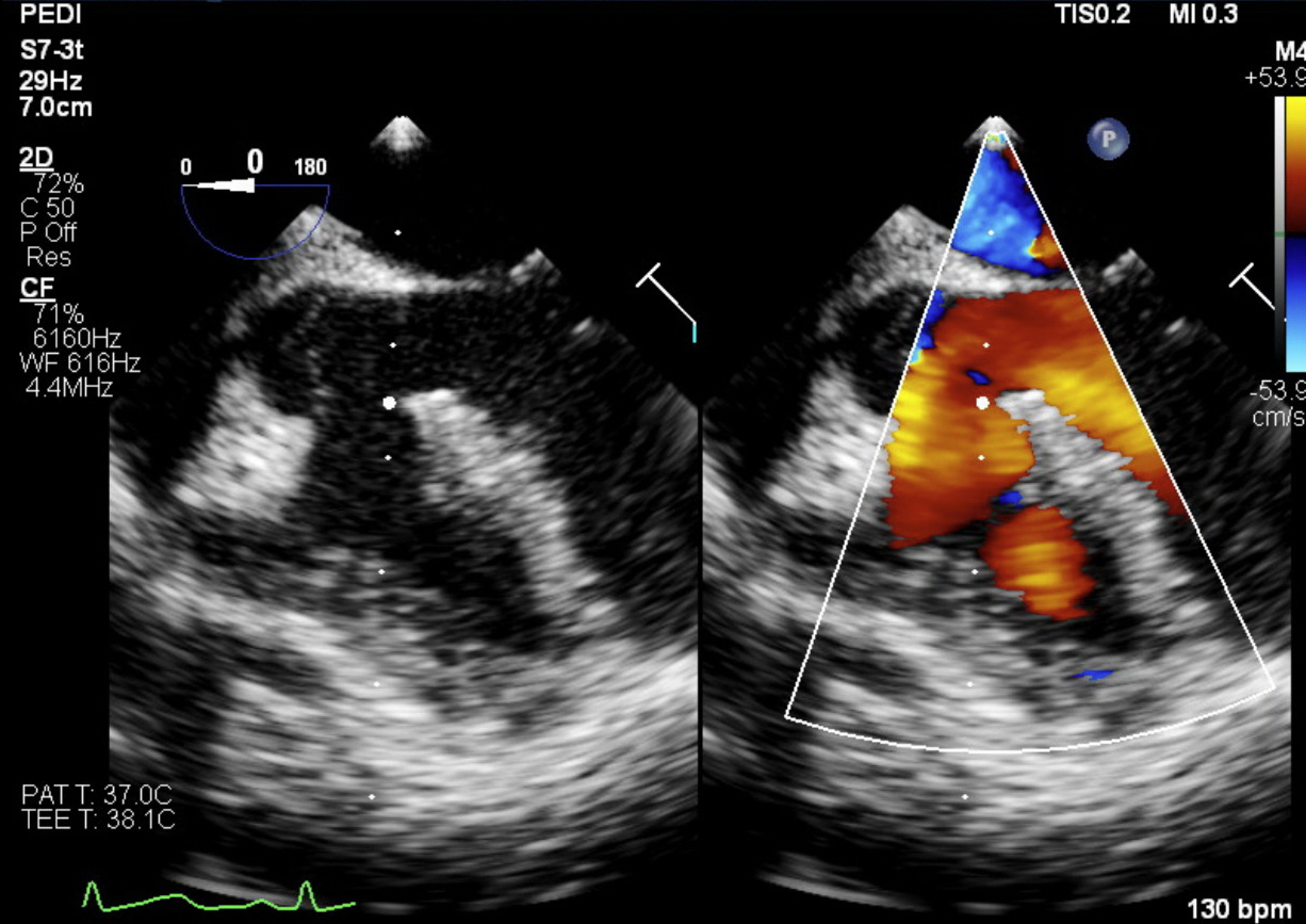
Image 2
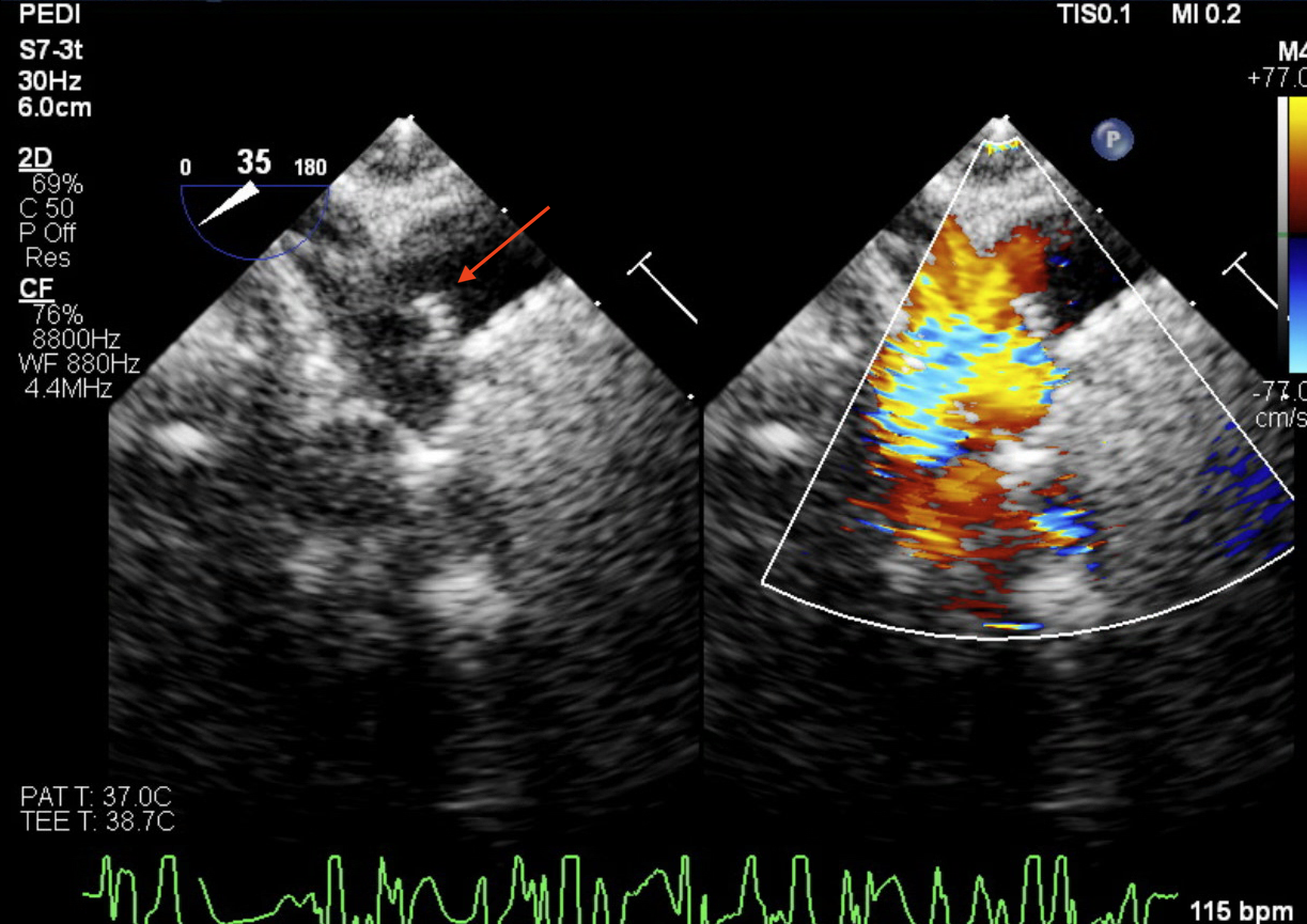
 

Image 3
Question 1

What view is demonstrated in Image 1 and which of the four characteristics of ToF can be seen?

Answer

Image 1 is a mid-esophageal 5 chamber view with color flow doppler color compare. To obtain this, a mid-esophageal 4 chamber view is obtained, omniplane from 0-10° and/or the probe can either be pulled up slightly or antero-flexed to visualize the more anterior aortic valve.

In this view, 3 of the 4 characteristics of Tetralogy of Fallot is demonstrated: Right ventricular hypertrophy, anterior malalignment VSD and over-riding aorta. The 4th, pulmonary stenosis is not seen in this view.

Question 2

In this midesophageal ascending aorta in short axis view with color flow doppler, the MPA, RPA and LPA are seen. What else can be seen in the left panel of this color compare, as indicated by the red arrow?

Answer

This is a mid-esophageal ascending aorta in short axis with color flow doppler color compare. The MPA is seen bifurcating to the RPA and LPA. Although not well seen in this image, this view will typically show the ascending aorta in short axis immediately left of the MPA as well as the SVC in short axis to the furthest left as depicted in the schematic image below.

The arrow is pointing at an echo dense structure which given the patient’s history is the PDA stent. It is seen partially protruding from the PDA into the bifurcation towards the LPA with color flow doppler showing some turbulent flow.  

Question 3

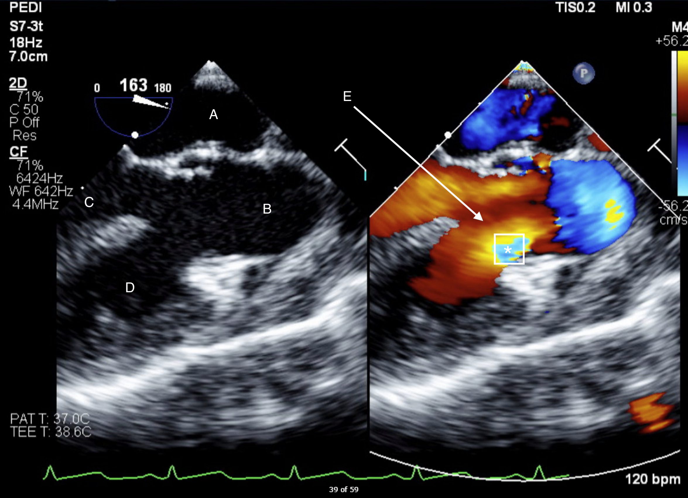
What view is shown in image 3? Identify the labels A through E.

Answer

Image 3 is a mid-esophageal long axis view with color flow doppler color compare. Usually, an omniplane angle of 120-140° is needed to obtain the view. The structures labeled are A) Left Atrium, B) Ascending aorta; since the image is clipped during systole, the aortic valve leaflets are open and not well visualized, C) Left ventricle/LV outflow tract, D) Right ventricle, and E) The arrow is pointing to the VSD which during systole is showing non-turbulent flow from the LV to the RV.

Additionally, there is some turbulence seen where the RVOT is expected (marked by the asterisk on the RV side of the VSD). This is the conal or infundibular septum. Normally, the conal septum fuses with the muscular interventricular septum and separates the Aortic and Pulmonary outflow tracts. In ToF the conal septum is pushed forward into the RV (anteriorly malaligned) resulting in obstruction to pulmonary blood flow.

Image 4

Question 4

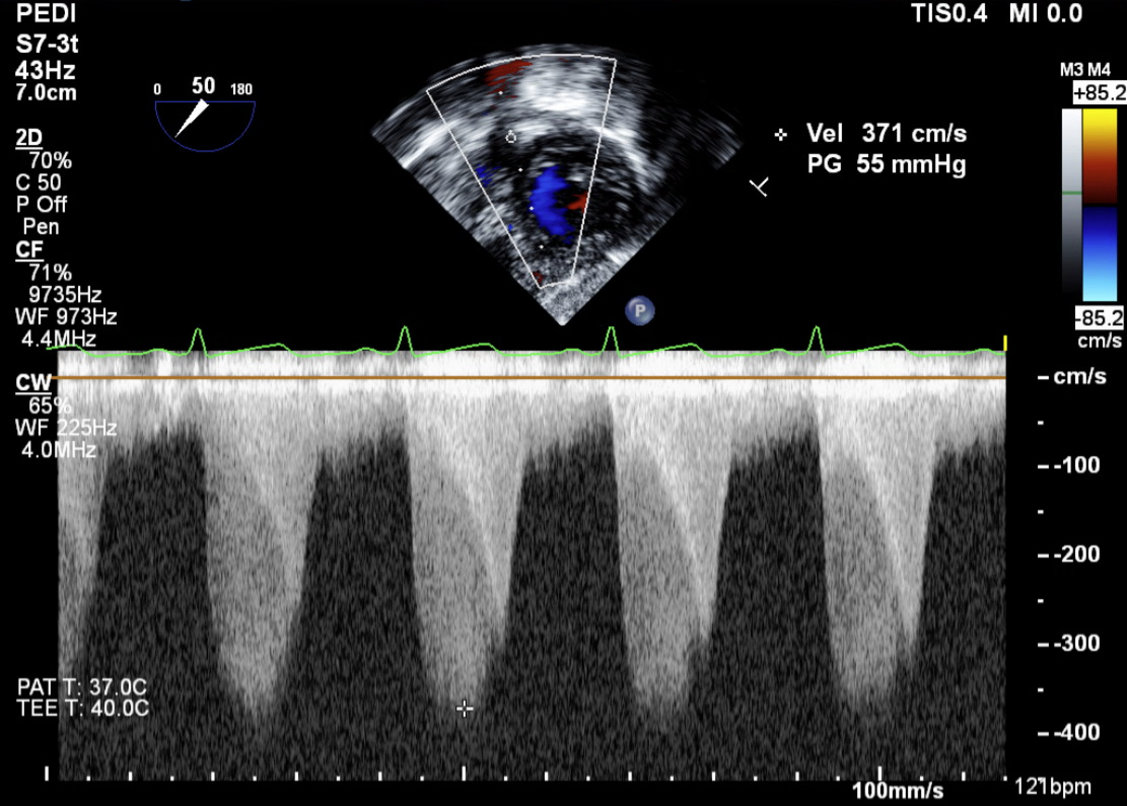
What view is used to obtain the spectral doppler in Image 4?

Answer

The view used to obtain this continuous wave doppler is a transgastric RV basal view, which is obtained at the transgastric level with some anteflexion and omniplane 0-20°. The image has been inverted (utilizing the up/down function) such that the ultrasound source is shown originating at the bottom and thus the pulmonary valve is expected near the top, as shown by the continuous wave cursor location.

Question 5

What doppler pattern is seen, and what does this demonstrate?

Answer

There are two continuous wave doppler waveforms shown here. The parabolic waveform represents a fixed obstruction consistent with pulmonary stenosis. The “sharp tooth” or “dagger” pattern also seen is characteristic of a dynamic obstruction occurring in the latter half of systole, suggestive of dynamic RVOT obstruction. A gradient of 55 mmHg coincides with moderate pulmonary stenosis. This patient had both dynamic RVOT obstruction as well as multilevel (sub-valvar and valvar pulmonary stenosis).

The patient undergoes Tetralogy of Fallot repair including RVOT muscle resection, VSD closure, pulmonary valvotomy, MPA and RVOT reconstruction and enlargement without a transannular patch. Separation from cardiopulmonary bypass was uneventful. Post bypass TEE shows the following:

Video 1

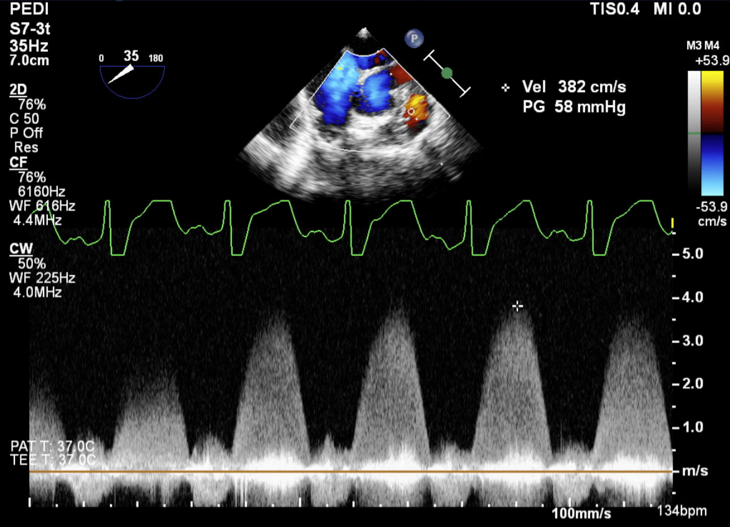
 Image 5


Question 6

Based on the video clip and Image 5, what residual lesion is shown?

Answer

The video clip is a deep transgastric RV outflow view which is obtained with omniplane 90°. The color flow doppler shows an unobstructed RVOT with turbulent flow at the level of the pulmonary valve annulus, despite MPA and RVOT reconstruction with appropriate sizing of the pulmonary valve with a 10mm probe/dilator. Although difficult to discern, pulmonary valve leaflet motion is seen but an enface view could not be obtained to assess the true leaflet morphology. The pulmonary valve is often difficult to assess with TEE due to it being the most anterior. Image 5 is a Mid-esophageal RV-inflow-outflow view. Once again high velocity flow at the level of the pulmonary valve is seen with a PG of 58mmHg which is similar to pre-operative values.

The decision is made to go back on cardiopulmonary bypass to repair the residual moderate pulmonary stenosis at the level of the pulmonary valve annulus. Due to the left anterior descending (LAD) artery course traversing immediately below the pulmonary valve/infundibular region, a transannular patch could not be performed to address the stenosis. Therefore, a 15mm aortic homograft was used as an RV-PA conduit and the post bypass echo clip is shown in Image 6.

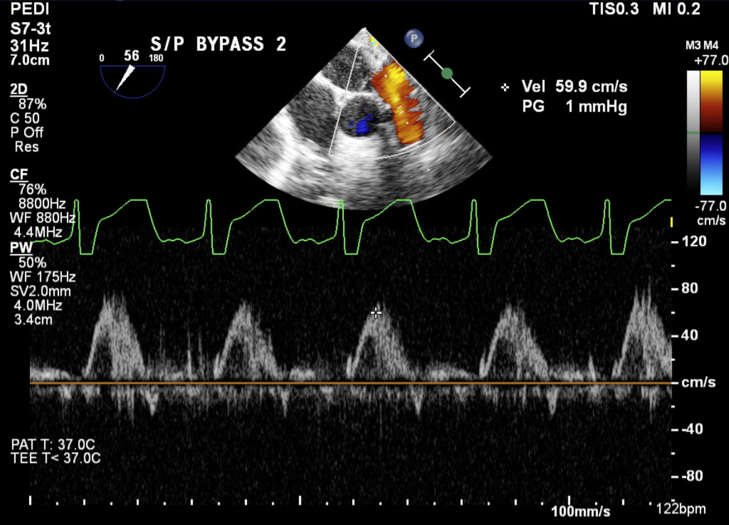
Image 6


Question 7

What view is used to obtain the pulse wave doppler in Image 6? What does a peak gradient of 1mmHg indicate?

Answer

This is also a mid-esophageal RV-inflow-outflow view with color flow doppler and pulse wave doppler revealing a peak gradient of 1mmHg (no obstruction or stenosis) after placement of an aortic homograft to serve as the RV-PA conduit. The bifurcation of the branch PAs and partial native LPA is seen with non-turbulent flow.

Discussion:

Tetralogy of Fallot (ToF) with PS is comprised of 4 major defects, as already mentioned above:

  • Anterior malalignment VSD with
  • Over-riding aorta over the muscular septum,
  • Right ventricular hypertrophy and possible RVOT narrowing/obstruction and,
  • Pulmonary stenosis consisting of any combination of subvalvar, valvar and supravalvar stenosis.


The spectrum of Tetralogy of Fallot also includes absent pulmonary valve and pulmonary atresia with and without major aortopulmonary collateral arteries.

Patients with ToF with PS can be asymptomatic (“Pink Tet”), have cyanotic Tet spells (with stress or agitation) or be cyanotic at baseline, depending on the degree of pulmonary blood-flow obstruction.

Modern day surgical repair usually consists of closing the VSD, alleviating the pulmonary obstruction and maintaining RV function for as long as possible. This is achieved by muscle bundle resection or RVOT resection and transannular patch, and if possible, valve sparing repair.(1,2) The degree of PS is dependent on the leaflet anatomy as well as degree of annular hypoplasia. It is often acceptable to have some degree of pulmonary insufficiency or even free PI often after transannular patch method is performed, which is well tolerated until the patient can undergo transcatheter pulmonary valve replacement (TPVR) or surgical replacement typically in adulthood. Valve sparing approach avoids a ventriculotomy which can contribute to post operative RV dysfunction, scarring and arrhythmias.

This patient presented with severe cyanosis in the neonatal period necessitating ductal stenting to ensure pulmonary blood flow. A transannular patch was not possible due to the course of the LAD. Pre-surgical imaging (TTE, TEE or cardiac CT) is often utilized to assess coronary anatomy in ToF, however the surgeon will ultimately make this decision upon visualization of the heart and corresponding coronary anatomy.

For a more in-depth discussion on the echo exam for Tetralogy of Fallot, please view the Echo tutorial here:  https://ccasociety.org/education/echoimage/intraoperative-evaluation-tetralogy/

The pre-operative transesophageal echocardiographic assessment should include the following:

  • Atrial septum for presence of an atrial communication
  • Right ventricular function
  • VSD size and location
  • Presence of any additional VSDs (that may be missed in the preop transthoracic exams)
  • Degree of RVOT obstruction, dynamic obstruction
  • Pulmonary valve size and morphology
  • MPA and branch PA size
  • Aortic valve and subaortic morphology
  • Coronary artery anatomy

The post-operative transesophageal echocardiographic assessment should focus on the following

  • Any atrial communication (often left intentionally), and directionality of flow across the communication
  • Tricuspid valve competency
  • Right ventricular pressure estimation if there is adequate tricuspid regurgitation jet
  • Right ventricular systolic function
  • Presence of any residual ventricular communication
  • PI, residual PS and branch PA flow
  • Left ventricular systolic function
  • Aortic valve morphology and competency

References:

  1.  Vanderlaan RD, Barron DJ. Optimal Surgical Management of Tetralogy of Fallot. CJC Pediatr Congenit Heart Dis. 2023 Sep 13;2(6Part A):352-360. doi: 10.1016/j.cjcpc.2023.09.003. PMID: 38161666; PMCID: PMC10755770.
  2. Miller JR, Stephens EH, Goldstone AB et al. The American Association for Thoracic Surgery (AATS) 2022 expert consensus document: Management of infants and neonates with tetralogy of Fallot. J Thorac Cardiovasc Surg 2023;165:221-50